
α-Carotene does not occupy the galectin-3 carbohydrate-recognition site in a blind docking screen benchmarked against olitigaltin
- Department of Biotechnology, Muthayammal College of Arts and Science (Autonomous),, Rasipuram, Namakkal, Tamilnadu, India
* Correspondence: ibtkavitha.d@gmail.com
Highlights
- Eight blueberry phytochemicals and olitigaltin were docked blind to galectin-3
- Olitigaltin located the carbohydrate-recognition site with no site restraint
- α-Carotene ranked best among the phytochemicals at −6.1 kcal/mol
- α-Carotene bound an adjacent hydrophobic patch and formed no hydrogen bonds
- Docking rank alone does not demonstrate galectin-3 target engagement
Abstract
Galectin-3 is a β-galactoside-binding lectin implicated in inflammation, fibrosis and adverse ventricular remodelling after myocardial infarction, and dietary phytochemicals are frequently screened against it by molecular docking. Such screens rarely include a reference inhibitor, and rarely verify where the predicted ligand actually binds. We docked eight phytochemicals reported from Vaccinium corymbosum L. (blueberry), together with the clinical-stage galectin-3 inhibitor olitigaltin (TD139), against the human galectin-3 carbohydrate-recognition domain (PDB 4BM8, 0.96 Å). A single grid enclosing the entire protein was used, with identical parameters for all nine ligands (AutoDock Vina via PyRx, exhaustiveness 32). Olitigaltin returned the most favourable score (−7.9 kcal/mol) and, without any site restraint, located the canonical carbohydrate-recognition site, forming hydrogen bonds with Asn160, Arg144 and Gly238 and π-interactions with His158, Arg162 and Trp181. α-Carotene ranked highest among the phytochemicals (−6.1 kcal/mol) but occupied an adjacent hydrophobic surface, contacting Pro117, Ile115, Tyr118 and Ala146 exclusively through alkyl and π-alkyl interactions and forming no hydrogen bonds. α-Carotene therefore does not engage the residues that determine galectin-3 carbohydrate
binding, and its docking rank should not be read as evidence of target engagement. Blind docking screens of phytochemicals should report a reference inhibitor and the identity of the contacted residues, not scores alone.
Keywords
1. Introduction
Myocardial infarction remains a leading cause of death worldwide, and the ventricular remodelling that follows ischaemic injury determines much of its long-term morbidity (Reed et al., 2017; World Health Organization, 2025). Galectin-3, a β-galactoside-binding lectin, is released by activated macrophages in the infarcted myocardium and promotes fibroblast activation, collagen deposition and adverse remodelling; genetic and pharmacological inhibition attenuates these processes in animal models (de Boer et al., 2010; Frangogiannis, 2014; Meijers et al., 2015; Wang et al., 2023; Yu et al., 2013). It is therefore an attractive target for small-molecule intervention.
Galectin-3 recognises its ligands through a shallow groove on the carbohydrate-recognition domain, in which a conserved network of residues — Arg144, His158, Asn160, Arg162, Asn174, Trp181 and Glu184 — forms hydrogen bonds and stacking contacts with the galactose ring. Inhibitor development has been directed almost exclusively at this groove, and the thiodigalactoside olitigaltin (TD139, GB0139) binds it with nanomolar affinity and has reached clinical evaluation (Hsieh et al., 2016). Because galectin-3 function depends on carbohydrate binding, a compound that does not occupy this groove has no evident route to inhibiting the protein.
Dietary phytochemicals are widely screened against galectin-3 and comparable targets by molecular docking, and carotenoids in particular are reported to have antioxidant and anti-inflammatory properties relevant to cardiovascular protection (Fiedor & Burda, 2014; Kaulmann & Bohn, 2014; Zhuang et al., 2022). Two weaknesses recur in these studies. First, scores are reported without a reference inhibitor, so the reader cannot judge whether a given value is meaningful. Second, when the search grid encloses the whole protein — a common and otherwise reasonable choice when the site is not assumed in advance — the reported pose is the global optimum anywhere on the surface, which need not be the functional site; the contacted residues are seldom examined. We therefore screened eight phytochemicals reported from Vaccinium corymbosum L. against galectin-3 under unbiased whole-protein conditions, with olitigaltin included as an internal reference under identical settings, and asked not only which compound scored best but where it bound.
2. Materials and methods
2.1 Protein and ligand preparation
The crystal structure of the human galectin-3 carbohydrate-recognition domain was obtained from the RCSB Protein Data Bank (Berman et al., 2000), entry 4BM8, solved by X-ray diffraction at 0.96 Å resolution (Noresson et al., 2014). Chain A was retained; water molecules and the co-crystallised ligand were removed, polar hydrogens were added and the structure was energy-minimised in BIOVIA Discovery Studio before conversion to PDBQT format.
Eight cardioprotective phytochemicals reported from Vaccinium corymbosum L. were identified using Dr. Duke's Phytochemical and Ethnobotanical Database. Their three-dimensional structures, and that of the reference inhibitor olitigaltin, were retrieved from PubChem (Kim et al., 2023), energy-minimised in PyRx using the universal force field and converted to PDBQT format (Dallakyan & Olson, 2015). Compound identities are given in Table 1.
2.2 Blind molecular docking
Docking was performed with AutoDock Vina 1.2.0 through the PyRx interface (Eberhardt et al., 2021; Trott & Olson, 2010). No binding site was assumed. A single search grid enclosing the entire protein was defined, centred at x = −12.32, y = 0.91, z = 5.34 with dimensions 41.55 × 45.15 × 40.31 Å, and the same grid centre, grid dimensions and search settings were applied to all nine ligands, including the reference inhibitor. The resulting search volume of approximately 7.6 × 10⁴ ų exceeds the 2.7 × 10⁴ ų threshold above which AutoDock Vina advises raising the exhaustiveness setting to compensate for the larger search; exhaustiveness was therefore set to 32, four times the program default. For each ligand the pose with the lowest predicted binding energy was retained and taken forward for interaction analysis.
2.3 Interaction and ADMET analysis
Protein–ligand contacts in the top-ranked poses were identified and classified in Discovery Studio Visualizer, and residue identities and contact distances were recorded. Physicochemical and pharmacokinetic descriptors were predicted using SwissADME (Daina et al., 2017) and acute oral toxicity using ProTox-3.0 (Banerjee et al., 2024).
3. Results and discussion
3.1 The reference inhibitor outscored every phytochemical
Predicted binding affinities are given in Table 1. Olitigaltin returned the most favourable value of the nine ligands, −7.9 kcal/mol. Among the phytochemicals, α-carotene ranked highest at −6.1 kcal/mol, followed by ferulic acid (−5.1 kcal/mol), with the remaining compounds between −4.4 and −3.1 kcal/mol. The 1.8 kcal/mol separation between olitigaltin and the best phytochemical is consistent with expectation: olitigaltin is the product of structure-guided optimisation against this groove, whereas the phytochemicals are dietary constituents under no selective pressure to bind it.
Molecular mass should be kept in view when reading this ranking. α-Carotene is the largest phytochemical in the set at 536.9 g/mol, and AutoDock Vina scores, which are not normalised for ligand size, tend to favour larger molecules through accumulated contact terms. Part of its advantage over the smaller phytochemicals, several of which are fragments of 74–194 g/mol, is therefore attributable to size rather than to complementarity.
Table 1. Identity and predicted binding affinity of the eight Vaccinium corymbosum phytochemicals and the reference inhibitor olitigaltin, ranked by docking score. All ligands were docked against PDB 4BM8 using an identical whole-protein grid.
Compound | PubChem CID | Molecular formula | Molecular weight (g/mol) | Binding affinity (kcal/mol) |
|---|---|---|---|---|
Olitigaltin (reference) | 73774610 | C₂₈H₃₀F₂N₆O₈S | 648.64 | −7.9 |
α-Carotene | 6419725 | C₄₀H₅₆ | 536.87 | −6.1 |
Ferulic acid | 445858 | C₁₀H₁₀O₄ | 194.18 | −5.1 |
Niacin | 938 | C₆H₅NO₂ | 123.11 | −4.4 |
Arginine | 6322 | C₆H₁₄N₄O₂ | 174.20 | −4.2 |
Nerol | 643820 | C₁₀H₁₈O | 154.25 | −4.2 |
Benzaldehyde | 240 | C₇H₆O | 106.12 | −4.0 |
Alanine | 5950 | C₃H₇NO₂ | 89.09 | −3.7 |
Butan-1-ol | 263 | C₄H₁₀O | 74.12 | −3.1 |
3.2 Only the reference inhibitor occupied the carbohydrate-recognition site
Because no site was specified, the position of each top-ranked pose is itself a result. Olitigaltin converged on the canonical carbohydrate-recognition groove (Figure 1A). Its contacts (Figure 2A) comprise conventional hydrogen bonds to Asn160 (2.85 Å), Arg144 (2.83 Å) and Gly238 (1.95 and 2.53 Å), a carbon–hydrogen bond to Asp239 (3.28 Å), π-cation interactions with His158 (3.82 Å) and Arg162 (3.67 Å), a π–π stacked interaction with Trp181 (5.45 Å), and halogen contacts to Asn174 (2.16 Å) and Glu184. These are the residues that define β-galactoside recognition in galectin-3, and their recovery from an unbiased whole-protein search, with distances typical of the thiodigalactoside series, is a stringent check that the docking protocol was able to identify the functional site when a true ligand was supplied.
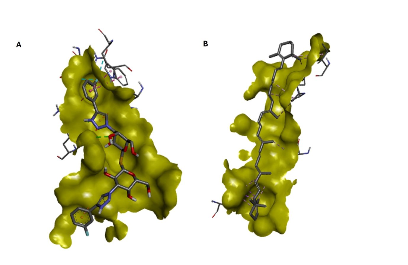
α-Carotene did not. Its top-ranked pose lay along an adjacent hydrophobic surface (Figure 1B), and its contacts (Figure 2B) consisted exclusively of alkyl and π-alkyl interactions with Pro117 (4.07 and 5.11 Å), Ile115 (4.37 and 4.86 Å), Tyr118 (4.45 and 4.60 Å) and Ala146 (4.42 Å), together with long-range contacts to Trp181 (4.74–7.01 Å) and His158 (4.72 Å). No hydrogen bond was formed. This is the expected consequence of its chemistry: α-carotene is a C₄₀ hydrocarbon with no hydrogen-bond donors or acceptors and no polar surface, and is constitutionally incapable of reproducing the hydrogen-bonding pattern through which galectin-3 recognises galactosides.
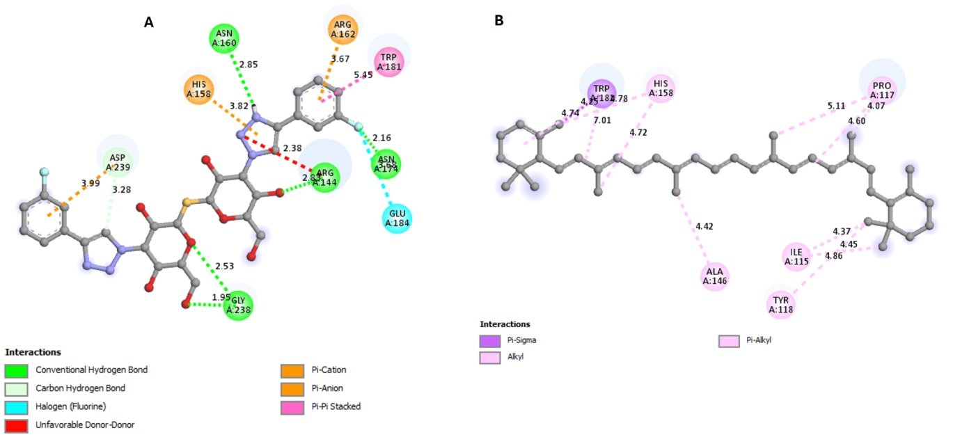
Of the nine residues contacted by olitigaltin, α-carotene shares only two, Trp181 and His158, and does so at 4.7 Å or more, which is peripheral rather than engaged. The two ligands are therefore not competing for the same site in any meaningful sense. Since galectin-3 exerts its profibrotic effects through carbohydrate-dependent cross-linking of glycoconjugates, a ligand that lies outside the recognition groove has no established mechanism by which to interfere with that function, whatever its docking score. The ranking in Table 1 and the binding-site assignment in Figure 2 thus support different conclusions, and the second is the more informative.
3.3 Predicted physicochemical and toxicity profiles
Predicted descriptors are summarised in Table 2. α-Carotene returned a consensus LogP of 8.27 with no hydrogen-bond donors or acceptors, a profile that lies far outside conventional oral drug-likeness criteria. This is consistent with the known behaviour of dietary carotenoids, whose absorption is low, highly variable and strongly dependent on food matrix and lipid co-ingestion (Nagao, 2014). In the BOILED-Egg analysis one compound fell outside the plotted range, and the remaining phytochemicals distributed across the regions associated with passive gastrointestinal absorption. ProTox-3.0 assigned α-carotene a predicted oral LD₅₀ of 1510 mg/kg and olitigaltin 1000 mg/kg, both corresponding to toxicity class IV; the prediction for α-carotene rested on a higher average structural similarity to the training set (71.45%) than that for olitigaltin (36.83%).
Table 2. Predicted physicochemical and pharmacokinetic descriptors (SwissADME). HBD, hydrogen-bond donors; HBA, hydrogen-bond acceptors; Log Kp, skin permeation coefficient; CYP, cytochrome P450 isoform predicted to be inhibited.
Compound | HBD | HBA | Consensus LogP | Log Kp (cm/s) | Aromatic heavy atoms | CYP inhibition |
|---|---|---|---|---|---|---|
Olitigaltin (reference) | 6 | 14 | 2.24 | −10.09 | 22 | CYP3A4 |
α-Carotene | 0 | 0 | 8.27 | 0.12 | 0 | None |
Ferulic acid | 2 | 4 | 1.41 | −6.41 | 6 | None |
Niacin | 1 | 3 | 0.47 | −6.80 | 6 | None |
Arginine | 4 | 4 | −2.99 | −10.34 | 0 | None |
Nerol | 1 | 1 | 2.92 | −4.71 | 0 | None |
Benzaldehyde | 0 | 1 | 1.43 | −5.90 | 6 | CYP1A2 |
Alanine | 2 | 3 | 0.42 | −8.95 | 0 | None |
Butan-1-ol | 1 | 1 | 1.69 | −6.13 | 0 | None |
These descriptors are secondary to the binding-site result. A favourable predicted toxicity class carries no weight if the compound does not engage the target, and the lipophilicity of α-carotene would in any case present a formulation problem before any pharmacological question could be asked.
3.4 Implications and limitations
The practical message is methodological. A screen reporting only that α-carotene achieved −6.1 kcal/mol against galectin-3 would have supported a claim of target engagement that the same data refute once the contacted residues are examined. Including a reference inhibitor under identical conditions cost one additional docking run and provided both a calibrated scale and a positive control on the search procedure; inspecting the contact residues cost nothing. We suggest both should be routine in phytochemical docking studies, and that where a whole-protein grid is used the site of the reported pose be stated explicitly.
Several limitations apply. The receptor was treated as rigid and a single crystallographic conformation was used, so induced fit was not modelled. Vina scores are not normalised for ligand size, and scores obtained at chemically different surface sites are not strictly comparable, since the scoring function was parameterised on ligands bound within defined pockets. Formal redocking of the co-crystallised 4BM8 ligand with calculation of root-mean-square deviation was not performed, although the independent recovery of the canonical site by olitigaltin provides comparable assurance. No molecular dynamics simulation, binding assay or cell-based experiment was undertaken, and the predicted ADMET values are unvalidated. The findings are computational and hypothesis-generating.
4. Conclusion
Under unbiased whole-protein docking with a reference inhibitor included at identical settings, olitigaltin located the galectin-3 carbohydrate-recognition site and reproduced its established contacts, whereas α-carotene — the best-scoring of eight Vaccinium corymbosum phytochemicals — bound an adjacent hydrophobic surface and formed no hydrogen bonds. α-Carotene is therefore not a galectin-3 carbohydrate-site ligand, and its rank in the screen reflects size and hydrophobic surface complementarity rather than target engagement. Docking screens of dietary compounds should report a reference inhibitor and the identity of contacted residues; scores alone can support conclusions that the underlying poses do not.
Data availability
Receptor and ligand PDBQT files, the docking configuration file and the output logs are available from the corresponding author on reasonable request.
References
- Banerjee, P., Kemmler, E., Dunkel, M., & Preissner, R. (2024). ProTox 3.0: A webserver for the prediction of toxicity of chemicals. Nucleic Acids Research, 52(W1), W513–W520. https://doi.org/10.1093/nar/gkae303
- Berman, H. M., Westbrook, J., Feng, Z., Gilliland, G., Bhat, T. N., Weissig, H., Shindyalov, I. N., & Bourne, P. E. (2000). The Protein Data Bank. Nucleic Acids Research, 28(1), 235–242. https://doi.org/10.1093/nar/28.1.235
- Daina, A., Michielin, O., & Zoete, V. (2017). SwissADME: A free web tool to evaluate pharmacokinetics, drug-likeness and medicinal chemistry friendliness of small molecules. Scientific Reports, 7, Article 42717. https://doi.org/10.1038/srep42717
- Dallakyan, S., & Olson, A. J. (2015). Small-molecule library screening by docking with PyRx. In J. E. Hempel, C. H. Williams, & C. C. Hong (Eds.), Chemical biology: Methods and protocols (Methods in Molecular Biology, Vol. 1263, pp. 243–250). Humana Press. https://doi.org/10.1007/978-1-4939-2269-7_19
- de Boer, R. A., Yu, L., & van Veldhuisen, D. J. (2010). Galectin-3 in cardiac remodeling and heart failure. Current Heart Failure Reports, 7(1), 1–8. https://doi.org/10.1007/s11897-010-0004-x
- Eberhardt, J., Santos-Martins, D., Tillack, A. F., & Forli, S. (2021). AutoDock Vina 1.2.0: New docking methods, expanded force field, and Python bindings. Journal of Chemical Information and Modeling, 61(8), 3891–3898. https://doi.org/10.1021/acs.jcim.1c00203
- Fiedor, J., & Burda, K. (2014). Potential role of carotenoids as antioxidants in human health and disease. Nutrients, 6(2), 466–488. https://doi.org/10.3390/nu6020466
- Frangogiannis, N. G. (2014). The inflammatory response in myocardial injury, repair, and remodelling. Nature Reviews Cardiology, 11(5), 255–265. https://doi.org/10.1038/nrcardio.2014.28
- Hsieh, T.-J., Lin, H.-Y., Tu, Z., Lin, T.-C., Wu, S.-C., Tseng, Y.-Y., Liu, F.-T., Hsu, S.-T. D., & Lin, C.-H. (2016). Dual thio-digalactoside-binding modes of human galectins as the structural basis for the design of potent and selective inhibitors. Scientific Reports, 6, Article 29457. https://doi.org/10.1038/srep29457
- Kaulmann, A., & Bohn, T. (2014). Carotenoids, inflammation, and oxidative stress: Implications of cellular signaling pathways and relation to chronic disease prevention. Nutrition Research, 34(11), 907–929. https://doi.org/10.1016/j.nutres.2014.07.010
- Kim, S., Chen, J., Cheng, T., Gindulyte, A., He, J., He, S., Li, Q., Shoemaker, B. A., Thiessen, P. A., Yu, B., Zaslavsky, L., Zhang, J., & Bolton, E. E. (2023). PubChem 2023 update. Nucleic Acids Research, 51(D1), D1373–D1380. https://doi.org/10.1093/nar/gkac956
- Meijers, W. C., van der Velde, A. R., Pascual-Figal, D. A., & de Boer, R. A. (2015). Galectin-3 and post-myocardial infarction cardiac remodeling. European Journal of Pharmacology, 763(Pt A), 115–121. https://doi.org/10.1016/j.ejphar.2015.06.025
- Nagao, A. (2014). Bioavailability of dietary carotenoids: Intestinal absorption and metabolism. Japan Agricultural Research Quarterly, 48(4), 385–391. https://doi.org/10.6090/jarq.48.385
- Noresson, A. L., Oberg, C. T., Engstrom, O., Hakansson, M., Logan, D. T., Leffler, H., & Nilsson, U. J. (2014). Galectin-3C in complex with bisamido-thiogalactoside derivate 3 (PDB ID 4BM8) [Data set]. RCSB Protein Data Bank. https://doi.org/10.2210/pdb4BM8/pdb
- Reed, G. W., Rossi, J. E., & Cannon, C. P. (2017). Acute myocardial infarction. The Lancet, 389(10065), 197–210. https://doi.org/10.1016/S0140-6736(16)30677-8
- Trott, O., & Olson, A. J. (2010). AutoDock Vina: Improving the speed and accuracy of docking with a new scoring function, efficient optimization, and multithreading. Journal of Computational Chemistry, 31(2), 455–461. https://doi.org/10.1002/jcc.21334
- Wang, X., Gaur, M., Mounzih, K., Rodriguez, H. J., Qiu, H., Chen, M., Yan, L., Cooper, B. A., Narayan, S., Derakhshandeh, R., Rao, P., Han, D. D., Nabavizadeh, P., Springer, M. L., & John, C. M. (2023). Inhibition of galectin-3 post-infarction impedes progressive fibrosis by regulating inflammatory profibrotic cascades. Cardiovascular Research, 119(15), 2536–2549. https://doi.org/10.1093/cvr/cvad116
- World Health Organization. (2025, July 31). Cardiovascular diseases (CVDs) [Fact sheet]. Retrieved October 13, 2026, from https://www.who.int/news-room/fact-sheets/detail/cardiovascular-diseases-(cvds)
- Yu, L., Ruifrok, W. P. T., Meissner, M., Bos, E. M., van Goor, H., Sanjabi, B., van der Harst, P., Pitt, B., Goldstein, I. J., Koerts, J. A., van Veldhuisen, D. J., Bank, R. A., van Gilst, W. H., Silljé, H. H. W., & de Boer, R. A. (2013). Genetic and pharmacological inhibition of galectin-3 prevents cardiac remodeling by interfering with myocardial fibrogenesis. Circulation: Heart Failure, 6(1), 107–117. https://doi.org/10.1161/CIRCHEARTFAILURE.112.971168
- Zhuang, C., Yuan, J., Du, Y., Zeng, J., Sun, Y., Wu, Y., Gao, X.-H., & Chen, H.-D. (2022). Effects of oral carotenoids on oxidative stress: A systematic review and meta-analysis of studies in the recent 20 years. Frontiers in Nutrition, 9, Article 754707. https://doi.org/10.3389/fnut.2022.754707
Acknowledgements
The authors thank Ms. Tamilbharathi, Program Lead, OriGene Biosolution, Salem, Tamil Nadu, India, for her guidance on the docking validation strategy, in particular the inclusion of a reference inhibitor docked under identical conditions, which substantially strengthened the interpretation of the results.
Funding
This research received no external funding.
Author information
Authors and Affiliations
Department of Biotechnology, Muthayammal College of Arts and Science (Autonomous),, Rasipuram, Namakkal, Tamilnadu, India
Anjali T & Kavitha D
Contributions
Anjali T: Data curation, Formal analysis, Investigation, Methodology, Visualization, Writing – original draft. Kavitha D: Conceptualization, Supervision, Validation, Writing – review & editing, Resources.
Corresponding author
Correspondence to Kavitha D (ibtkavitha.d@gmail.com).
Ethics declarations
Ethics approval and consent to participate
Not applicable.
Competing interests
The authors declare that they have no conflict of interest.
Use of generative AI
Generative AI tools were not used in the design, analysis or interpretation of this study.
Additional information
Publisher’s note
OriGene Academic Press remains neutral with regard to jurisdictional claims in published maps and institutional affiliations.
Rights and permissions
Open Access This article is licensed under a Creative Commons Attribution 4.0 International License (CC BY 4.0), which permits use, sharing, adaptation, distribution and reproduction in any medium or format, provided appropriate credit is given to the original author(s) and the source, a link to the licence is provided, and any changes are indicated. Images or other third-party material in this article are included in the article’s licence unless stated otherwise in a credit line to the material. © 2026 The Author(s).
About this article
Cite this article
T A, D K. α-Carotene does not occupy the galectin-3 carbohydrate-recognition site in a blind docking screen benchmarked against olitigaltin. Int J Adv Interdis Res. 2026;6(2):e006.
Download citation (RIS) · BibTeX ·
- Received03 Sep 2026
- Revised20 Oct 2026
- Accepted21 Oct 2026
- Published24 Oct 2026
- DOIregistration in progress
Share this article
Anyone you share the following link with will be able to read this article: